Peptide Solubility Troubleshooting: A Researcher’s Guide
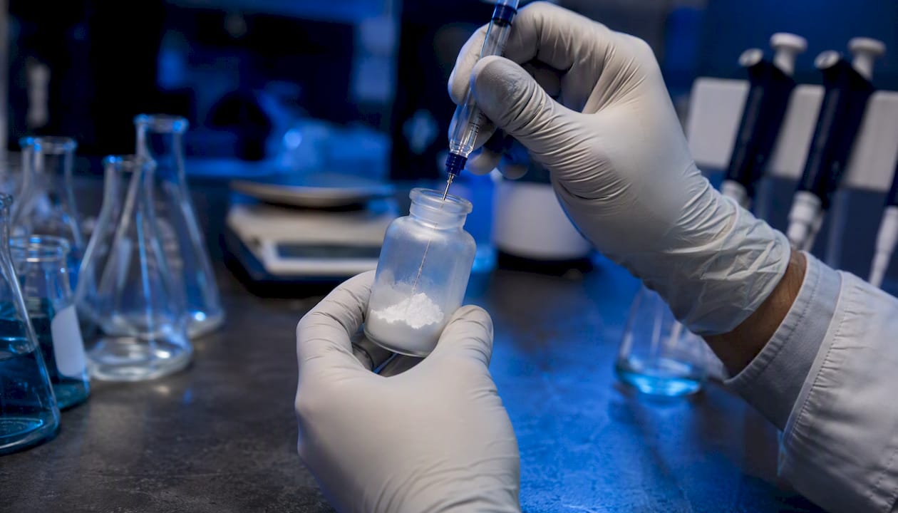
When a peptide won’t dissolve, the fastest path forward is a prioritized three-step sequence: try water or aqueous buffer first, then shift pH with dilute acid or base, then wet the pellet with a small volume of DMSO or acetonitrile before slow aqueous dilution. Most peptide solubility issues resolve at one of those three stages without requiring chaotropes or detergents that compromise downstream assays.
Before reaching for any solvent, run through this bench checklist:
Allow the lyophilized vial and your diluent to equilibrate to room temperature for 15–20 minutes before opening.
Work with a small test aliquot (0.1–0.2 mg) rather than the full vial.
Try sterile water or phosphate-buffered saline first; wait 5–10 minutes with gentle agitation.
If cloudy, assess net charge at pH 7: basic peptides (net positive) respond to 0.1% acetic acid; acidic peptides (net negative) respond to dilute NH4HCO3 or NH4OH.
If still insoluble, wet the pellet with 50–100 µL DMSO per mg, then dilute slowly into aqueous buffer.
Verify with a brief centrifugation (14,000 × g, 5 minutes) and visual clarity check before proceeding.
Note any cysteine or methionine residues: use degassed, acidic solvents and avoid oxidizing conditions.
Pro Tip: Never add cold solvent to a cold vial. Temperature mismatch is one of the most common causes of localized aggregation that resists even strong solubilizers — a failure mode that is entirely avoidable.
Key Takeaways
Effective peptide solubility troubleshooting follows a minimal-intervention sequence: water first, then pH adjustment, then small-volume organic co-solvent, with verification at each step before escalating.
Point Details Start minimal, escalate deliberately Try water at 1–2 mg/mL first; move to pH adjustment, then DMSO wetting only after each step fails. Verify before proceeding Centrifuge at 14,000 × g for 5–10 minutes and check A280 before assuming a clear solution is fully dissolved. Equilibrate temperature first Allow both the vial and diluent to reach room temperature (15–20 minutes) before any reconstitution attempt. Aliquot on first reconstitution Divide stock into single-use aliquots and freeze at −80°C immediately to prevent freeze–thaw degradation. Review COA before each new lot Batch-specific solvent recommendations and purity data on the COA should guide reconstitution strategy for every new lot.
Table of Contents
Why do peptides resist dissolution?
Peptide solubility troubleshooting: a stepwise bench protocol
Which solvents and reagents should you use?
Physical techniques that assist dissolution
How do you confirm a peptide is truly in solution?
Storage and handling for lyophilized and reconstituted peptides
Difficult sequences: amyloidogenic, hydrophobic, and cysteine-rich peptides
Can computational tools predict peptide solubility before you run the experiment?
Quick-reference cheat sheet: starting solvents by peptide class
Editorial perspective: what the protocol misses in practice
Sourcing peptides with reliable COA and batch documentation
Sources
Why do peptides resist dissolution?
Poor solubility is almost always traceable to one or more of four sequence-level factors: high hydrophobicity, unfavorable net charge at the working pH, length beyond roughly 25 residues, or a propensity to form beta-sheet secondary structure or amyloid-like aggregates. Understanding which factor dominates determines which troubleshooting branch to take first.
The isoelectric point (pI) is the pH at which a peptide carries zero net charge. At or near the pI, electrostatic repulsion between molecules drops to a minimum, and intermolecular attractive forces drive aggregation. Practically, this means a peptide that appears soluble at pH 4 may crash out of solution when the pH is adjusted toward physiological range. Knowing the calculated pI before reconstitution lets you choose a starting pH that keeps the peptide charged and repulsive enough to stay dispersed.
Hydrophobicity compounds the problem. Peptides with more than 50% hydrophobic residues, or with long uninterrupted hydrophobic stretches, tend to self-associate through van der Waals and hydrophobic interactions regardless of charge state. Transmembrane segments are the extreme case, but shorter sequences with multiple leucine, isoleucine, valine, or phenylalanine residues behave similarly. As LifeTein’s troubleshooting guide notes, hydrophobicity, charge mismatch, and sequence length are the three most frequent causes of failed dissolution.
Impurities and lyophilization residuals add a practical layer on top of the sequence-level factors. Residual TFA from HPLC purification, residual salts from synthesis buffers, and oxidized cysteine or methionine residues all reduce apparent solubility. A peptide that dissolved cleanly from a previous batch may resist dissolution from a new batch if the lyophilization cycle left different excipient levels or if oxidation occurred during shipping. This is why reviewing the Certificate of Analysis (COA) before reconstitution is a non-negotiable first step, not an afterthought.
A practical rule of thumb for initial solvent selection:
Net positive charge at pH 7 (more Arg, Lys, His than Asp, Glu): start with dilute acetic acid (0.1–1%).
Net negative charge at pH 7 (more Asp, Glu than basic residues): start with dilute NH4HCO3 or NH4OH (0.1%).
Near-zero net charge or high hydrophobicity: start with a small volume of DMSO or ACN as a wetting step before aqueous dilution.
Peptide solubility is not a fixed property — it is a function of pH, ionic strength, temperature, concentration, and the history of the sample. A peptide that “doesn’t dissolve” under one set of conditions may dissolve readily when even one of those variables is adjusted. The goal of troubleshooting is to identify which variable is the limiting factor, not to find a universal solvent.
The aggregation risk is sequence-specific and can be compounded by concentration. At high stock concentrations, even moderately hydrophobic peptides can nucleate aggregates that persist after dilution. Starting at 1–2 mg/mL rather than pushing toward 10 mg/mL is a practical hedge against this. A peer-reviewed review on peptide aggregation and dissolution strategies confirms that no single solubilization method works across all difficult sequences, and that amyloidogenic motifs in particular require case-by-case optimization.
For peptide formulation interactions that go beyond simple reconstitution, the formulation base itself can shift the effective solubility window significantly.
Peptide solubility troubleshooting: a stepwise bench protocol
The protocol below is designed to conserve material, preserve assay compatibility, and give clear decision points at each stage. Always start with a small test aliquot.
Equilibrate. Allow the sealed vial and your chosen diluent to reach room temperature (15–20 minutes). Do not skip this step.
Weigh a test aliquot. Work with 0.1–0.2 mg. Do not open the full vial until the reconstitution strategy is confirmed.
Add sterile water or PBS. Target 1–2 mg/mL. Vortex gently for 30 seconds, then let stand for 5 minutes. Clear solution: proceed to dilution and use. Cloudy but no visible particles: try gentle sonication (30–60 seconds in a bath sonicator at room temperature) and re-check. Visible particulate or gel: move to step 4.
Clear solution: proceed to dilution and use.
Cloudy but no visible particles: try gentle sonication (30–60 seconds in a bath sonicator at room temperature) and re-check.
Visible particulate or gel: move to step 4.
Adjust pH based on net charge. Basic peptide (net positive): add 0.1–1% acetic acid; try 10–30% acetic acid for strongly hydrophobic basic peptides per GenScript’s solubility guidelines.
Basic peptide (net positive): add 0.1–1% acetic acid; try 10–30% acetic acid for strongly hydrophobic basic peptides per GenScript’s solubility guidelines.
Acidic peptide (net negative): add 0.1% NH4OH or 10 mM NH4HCO3 dropwise. Neutral/hydrophobic: skip to step 5. Wait 5–10 minutes with gentle rolling agitation. Re-check clarity.
Neutral/hydrophobic: skip to step 5.
Wait 5–10 minutes with gentle rolling agitation. Re-check clarity.
Organic co-solvent wetting step. Add 50–100 µL DMSO per mg of peptide directly to the dry pellet (or to the cloudy suspension). Vortex until the pellet is fully wetted. Then dilute slowly into aqueous buffer, adding buffer dropwise while vortexing. Final DMSO fraction should stay below 10% for most cell assays, below 1% for sensitive enzymatic assays.
Sonication. If still cloudy after step 5, place the tube in a bath sonicator for 1–2 minutes at room temperature. Probe sonication is an option for small volumes but risks peptide degradation at high power settings.
Centrifuge and assess. Spin at 14,000 × g for 5–10 minutes. If a pellet forms, the supernatant concentration is lower than intended — measure A280 or use a Bradford assay to quantify. Discard the pellet or attempt re-solubilization with a stronger approach.
Escalate to chaotropes or detergents only if steps 1–7 fail. Urea (4–8 M) or guanidine HCl (4–6 M) can dissolve strongly hydrophobic or aggregated peptides but are incompatible with most cell-based and many enzymatic assays. If chaotropes are used, plan for dialysis or buffer exchange before downstream use.
Stop and contact the supplier if: the pellet persists after chaotrope treatment, the solution color is abnormal (yellow/brown suggesting oxidation or degradation), or the COA indicates a purity below 90% — these are signals of a batch quality issue rather than a reconstitution technique problem.
Key decision points at each stage:
Cloudy after water: likely near-pI aggregation or mild hydrophobicity — pH adjustment usually resolves it.
Particulate after pH adjustment: likely high hydrophobicity or amyloid nucleation — organic co-solvent wetting is the next step.
Pellet after centrifugation post-DMSO: likely irreversible aggregation or insoluble impurity — escalate to chaotropes or return to supplier.
Abnormal color or odor: stop and review COA; do not use the material.
The Sigma-Aldrich solubility guidelines recommend testing small aliquots and note that organic solvents should be removed by lyophilization if they are incompatible with downstream assays. The Thermo Fisher handling guide reinforces consulting the COA for batch-specific solvent recommendations before committing to any reconstitution strategy.
Which solvents and reagents should you use?
No universal solvent exists for peptide reconstitution. The right choice depends on the peptide’s sequence class, the target concentration, and what the downstream assay can tolerate. The table below maps common peptide types to recommended starting solvents and compatibility notes.
Peptide class First solvent Second solvent Assay compatibility note Charged (net positive, basic) 0.1–1% acetic acid in water 10–30% acetic acid Compatible with most assays; volatile, can be lyophilized away Charged (net negative, acidic) 0.1% NH4OH or 10 mM NH4HCO3 PBS pH 7 Compatible; NH4HCO3 is volatile and MS-compatible Hydrophobic (>50% hydrophobic residues) DMSO (50–100 µL/mg wetting) ACN/water (1:1), then dilute DMSO: keep <10% for cell assays, <1% for enzyme assays Neutral/mixed Water or PBS 0.1% acetic acid or DMSO Test water first; escalate based on result Cysteine-rich Degassed 0.1% acetic acid DMSO (if needed) Avoid oxidizing conditions; use degassed solvents throughout Amyloidogenic HFIP or DMSO monomerization Dilute into aqueous buffer HFIP is toxic; handle in fume hood; remove before cell assays
Specific reagent notes:
Acetic acid (0.1–30% in water): Preferred for basic peptides. Volatile, so it can be removed by lyophilization if needed. Does not interfere with most UV-based assays.
NH4OH (0.1% in water): Preferred for acidic peptides. Also volatile. Avoid concentrations above 0.5% — higher concentrations can cause asparagine/glutamine deamidation over time.
DMSO: The most widely used organic co-solvent. Effective wetting agent at 50–100 µL per mg. Penetrates cell membranes at high concentrations, which can confound cell-based assays. Keep final DMSO below 10% for most applications, and below 0.5–1% for sensitive cell viability assays.
ACN (acetonitrile): Useful for strongly hydrophobic peptides. More volatile than DMSO, which simplifies removal. Incompatible with many protein-binding assays at concentrations above 5%.
DMF (dimethylformamide): Higher solvating power than DMSO for some hydrophobic sequences, but more toxic and harder to remove. Use only when DMSO fails.
Urea (4–8 M) and guanidine HCl (4–6 M): Chaotropes that disrupt hydrogen bonding and hydrophobic interactions. Effective for strongly aggregated or gelling peptides. Incompatible with most cell assays and many enzymatic assays; require dialysis or desalting before use.
Mild detergents (SDS, Tween-20): Last resort for membrane-associated peptides. SDS is incompatible with native PAGE and most binding assays. Tween-20 at 0.01–0.1% is gentler but still affects membrane assays.
Safety and waste handling: DMSO, DMF, and ACN are absorbed through skin. Use nitrile gloves and work in a ventilated area. Guanidine HCl waste requires neutralization before disposal per institutional guidelines. HFIP (hexafluoroisopropanol, used for amyloidogenic peptides) is acutely toxic and must be handled in a chemical fume hood with appropriate PPE.
As Merck Millipore’s handling protocol notes, when using organic solvents, select an initial solvent that can be removed if needed — this preserves the option to reformulate without discarding the batch.
Physical techniques that assist dissolution
The most reliable physical assists are sonication, controlled warming, correct order of addition, and slow dilution. Each addresses a different failure mode, and applying them in the wrong order can make solubility worse.
Order of addition is the single most consequential variable. Dissolve the peptide completely in the minimum volume of organic co-solvent first, then add aqueous buffer dropwise while vortexing. Reversing this sequence — adding organic solvent to an aqueous suspension — creates transient high-organic zones that force localized precipitation, often producing aggregates that resist further solubilization. The Merck Millipore protocol is explicit on this point: complete dissolution in the organic phase before any aqueous addition.
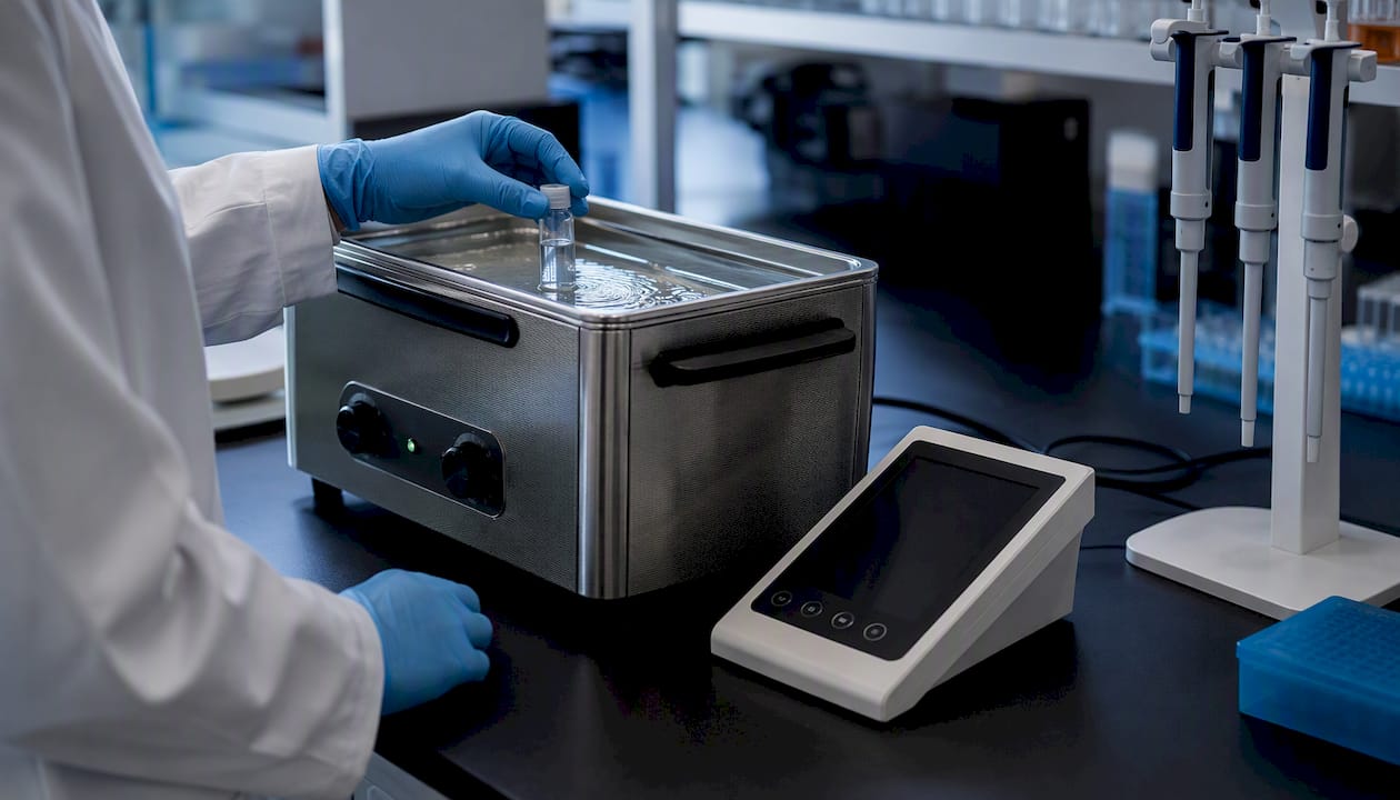
Sonication is effective for breaking up loose aggregates and hydrating dry pellets. A bath sonicator at room temperature for 30–120 seconds is the standard approach. Probe sonication delivers more energy but risks peptide fragmentation or oxidation at high amplitudes — keep probe sonication below 20% amplitude and limit to 30-second pulses with cooling intervals. Never sonicate cysteine-rich peptides without degassed solvent; cavitation introduces dissolved oxygen that accelerates disulfide scrambling.
Controlled warming (37–50°C water bath for 5–10 minutes) helps dissolve hydrophobic peptides that are kinetically trapped rather than thermodynamically insoluble. Warming reduces viscosity and increases molecular motion, which can break weak hydrophobic contacts. Do not exceed 50°C for peptides containing glutamine, asparagine, or methionine — deamidation and oxidation accelerate above that threshold.
Gentle rolling agitation (on a tube roller at 10–20 rpm) is preferable to vigorous vortexing for peptides prone to foaming or mechanical shear-induced aggregation. Vortexing is fine for most peptides but can introduce air bubbles that nucleate aggregation in amphipathic sequences.
Slow dilution: add aqueous buffer at no more than 10× the organic volume per addition step, waiting 30 seconds between additions.
Centrifugation (14,000 × g, 5–10 minutes) after each major step clarifies the solution and lets you assess whether a pellet is forming.
Filtration (0.22 µm PVDF or PES membrane) removes insoluble particulates but also removes any peptide that has aggregated — weigh the filtrate concentration by A280 or Bradford to account for losses.
Pro Tip: The most common non-obvious failure is adding cold buffer directly from the refrigerator to a peptide vial that was also stored cold. Both the vial and the diluent must be at room temperature before contact. Biostrata Research’s reconstitution guide identifies temperature mismatch as a top recurring failure mode — one that is easy to prevent and frequently overlooked.
How do you confirm a peptide is truly in solution?
The fastest practical verification sequence is: visual clarity check → brief centrifugation → UV absorbance at A280 (or A205 for peptides lacking aromatic residues). Escalate to dynamic light scattering (DLS) or size-exclusion chromatography (SEC) when subvisible aggregates are suspected or when assay results are inconsistent with expected concentration.
Visual clarity is a necessary but insufficient check. A solution can appear clear while containing subvisible aggregates in the 100 nm–1 µm range. Conversely, slight opalescence does not always indicate insoluble material — some peptide solutions are inherently slightly turbid at high concentrations.
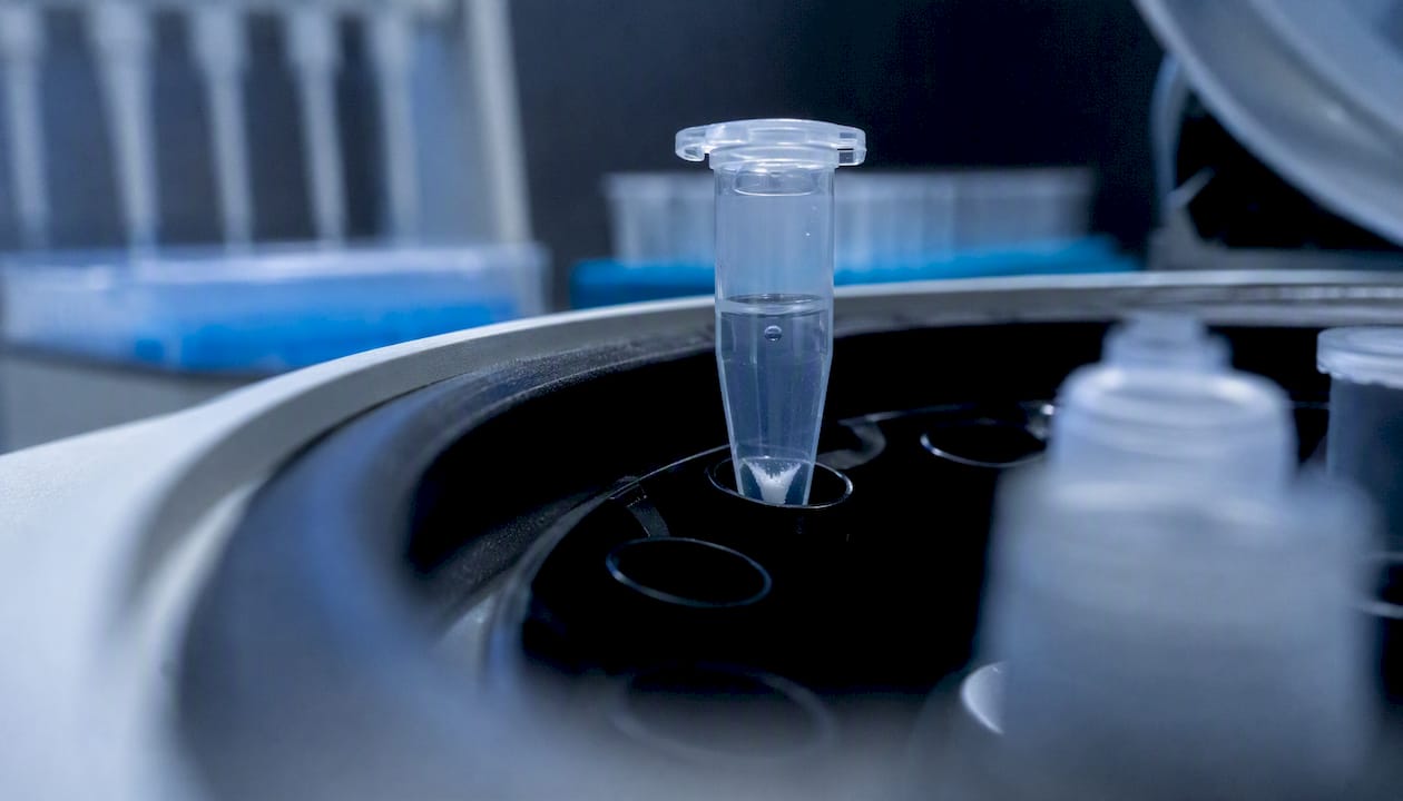
Centrifugation at 14,000 × g for 5–10 minutes is the most practical first escalation. A visible pellet after centrifugation confirms insoluble material. Measure A280 in the supernatant and compare to the expected absorbance for the target concentration (using the calculated molar extinction coefficient from the sequence). A significant drop from expected absorbance indicates that a fraction of the peptide is in the pellet.
Verification method What it detects Practical limit When to use Visual inspection Gross turbidity, particles >1 µm Cannot detect subvisible aggregates Always, as a first pass Centrifugation + A280 Insoluble fraction, approximate concentration Requires aromatic residues for A280; use A205 otherwise After each major reconstitution step DLS (dynamic light scattering) Particle size distribution, aggregates 1 nm–10 µm Requires DLS instrument; sensitive to dust When subvisible aggregation is suspected SEC (size-exclusion chromatography) Monomer vs. oligomer/aggregate fractions Requires HPLC setup; dilution during run When monomer purity is critical for assay Bradford/BCA assay Total soluble protein/peptide concentration Bradford underestimates some peptides When A280 is unreliable (no Trp/Tyr)
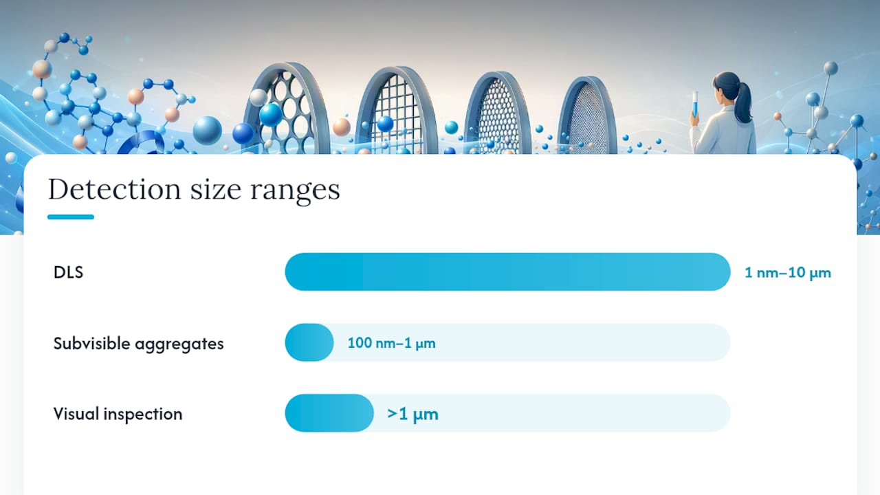
DLS is particularly useful for amyloidogenic peptides, where the solution can appear clear while containing nanometer-scale oligomers that are biologically active and assay-confounding. SEC provides the most definitive picture of monomer vs. aggregate distribution but requires an appropriate column and running buffer that does not itself alter aggregation state.
When to contact the supplier: if centrifugation consistently yields a pellet despite correct reconstitution technique, if A280 readings are far below expected (suggesting low actual purity), or if the COA purity is below 90%, request a new COA or a replacement batch. Batch-to-batch impurities and residual salts from synthesis can change apparent solubility in ways that no reconstitution technique can fully compensate for.
Storage and handling for lyophilized and reconstituted peptides
The most important storage rules are: keep lyophilized peptides at −20°C (or −80°C for long-term storage of sensitive sequences), aliquot reconstituted solutions before freezing, and avoid repeated freeze–thaw cycles. Each freeze–thaw cycle can reduce effective solubility by promoting aggregation nuclei that persist after re-thawing.
Stock concentration guidance varies by peptide class. Most standard peptides can be prepared at 1–5 mg/mL in appropriate solvent without solubility issues. Hydrophobic peptides are often limited to 0.5–1 mg/mL in aqueous systems; pushing higher concentrations forces aggregation that is difficult to reverse. Cysteine-rich peptides should be stored in degassed solvent at low concentration to minimize disulfide scrambling.
Vialing and residual excipients matter more than most researchers expect. Lyophilized peptides often contain residual TFA, acetate, or other counterions from the purification process. These affect both solubility and downstream assay compatibility. The Thermo Fisher handling guide stresses reviewing the COA for batch-specific solvent recommendations — a practice that should precede every reconstitution, not just the first one.
Storage format Recommended conditions Typical stability Key precautions Lyophilized (dry powder) −20°C, desiccated, sealed under inert gas 1–3 years (sequence-dependent) Equilibrate to room temperature before opening; avoid moisture Short-term solution stock 4°C, 1–7 days Days to 1 week Use within 1 week; keep sterile; avoid light for photosensitive sequences Long-term frozen stock −80°C, single-use aliquots Months to 1 year Aliquot before first freeze; never refreeze after thaw
Handling during receipt: inspect the vial for visible moisture or discoloration before opening. A yellow or brown tint in a lyophilized peptide suggests oxidation during shipping or storage. Review the COA for purity, counterion, and recommended solvent — this information is batch-specific and should not be assumed from a previous lot.
Pro Tip: Prepare single-use aliquots at the time of first reconstitution. Divide the stock into volumes that match your typical experiment size, freeze immediately at −80°C, and thaw only what you need. This workflow eliminates freeze–thaw degradation and makes it straightforward to trace any solubility change back to a specific aliquot or batch.
For a detailed walkthrough of lyophilization and reconstitution workflows, including order-of-operations and COA interpretation, that resource covers the full process from vial receipt to working stock preparation.
Difficult sequences: amyloidogenic, hydrophobic, and cysteine-rich peptides
These sequence classes need stronger, sequence-specific strategies. Standard reconstitution protocols often fail entirely, and attempting them without modification risks irreversible aggregation that wastes the entire vial.
Amyloidogenic peptides (e.g., Aβ fragments, IAPP, insulin fragments)
Amyloidogenic sequences nucleate beta-sheet aggregates rapidly in aqueous solution, often within minutes of reconstitution. The standard approach is monomerization before aqueous dilution. Dissolve the lyophilized peptide in neat HFIP (hexafluoroisopropanol) at 1–2 mg/mL, sonicate briefly, then aliquot and evaporate the HFIP under a gentle nitrogen stream or in a SpeedVac. The resulting thin film can then be dissolved in DMSO (5–10 µL per aliquot) and diluted into cold aqueous buffer immediately before use. The PubMed review on peptide aggregation and dissolution covers monomerization strategies for amyloidogenic sequences in detail and emphasizes that irreversible aggregation, once established, cannot be reversed by dilution or pH adjustment.
Warning: HFIP is acutely toxic and corrosive. Handle exclusively in a chemical fume hood with appropriate PPE. Never use HFIP in cell assays without complete removal.
Transmembrane and strongly hydrophobic peptides
These sequences often require neat DMSO or DMF as the primary solvent, with aqueous dilution kept to the minimum needed for the assay. Warming to 37–50°C during the initial dissolution step helps. For HPLC analysis, ACN/water gradients with 0.1% TFA are standard. If the peptide gels in aqueous buffer, consider 4–8 M urea as a chaotrope, with downstream dialysis. Sequence redesign (adding charged flanking residues) or encapsulation strategies may be necessary for sequences that cannot be adequately solubilized by any solvent combination.
Cysteine-rich and disulfide-containing peptides
Free cysteine residues oxidize readily in air, forming intermolecular disulfide bonds that cause irreversible aggregation. Reconstitute in degassed 0.1% acetic acid or degassed water, working quickly under nitrogen or argon if possible. Add a reducing agent (DTT at 1–5 mM, or TCEP at 0.5–1 mM) if the assay tolerates it. TCEP is preferred because it is more stable and does not interfere with most downstream assays. Avoid DMSO as the primary solvent for cysteine-rich peptides — DMSO can oxidize free thiols under some conditions.
Recognize irreversible aggregation by: persistent pellet after chaotrope treatment, gel-like consistency that does not respond to heating or sonication, and A280 readings far below expected even after extended solubilization attempts.
When irreversible aggregation is confirmed, lyophilize back to dry (if the peptide is still functional) and restart with a modified protocol, or contact the supplier for solubility testing data.
Pro Tip: For any sequence with three or more cysteine residues, request the COA’s disulfide bond map before reconstitution. Knowing whether the cysteines are free, protected, or already oxidized changes the entire solubilization strategy.
Can computational tools predict peptide solubility before you run the experiment?
In silico predictors can meaningfully reduce wasted material by ranking candidate sequences for relative solubility before synthesis or reconstitution, but they do not replace experimental validation for absolute solubility measurements.
CamSol-PTM, published in Nature Communications, extends the original CamSol intrinsic solubility predictor to peptides containing noncanonical amino acids. The authors report moderate-to-high Pearson correlation coefficients between predicted and experimentally measured relative solubilities across their validation sets, with performance varying by peptide series. The key word is “relative” — CamSol-PTM ranks sequences against each other, which is useful for lead optimization, but it does not output an absolute solubility value in mg/mL. The CamSol-PTM paper is the primary reference for understanding both the method’s capabilities and its dataset-specific limitations.
Computational solubility prediction is most valuable at the design stage, when you can still modify the sequence. Once a peptide is synthesized, the predictor’s main utility is helping you understand why it is difficult and which solvent class is most likely to work — not guaranteeing a solution.
A practical workflow for using in silico tools:
Input the sequence into CamSol-PTM or a comparable predictor (e.g., SOLpro, PepFold-based tools) before synthesis.
Flag sequences with predicted low relative solubility for sequence modification (adding charged residues, shortening hydrophobic stretches) or for planning a more aggressive reconstitution strategy.
After synthesis, run a small-scale solubility assay (PEG precipitation, ammonium sulfate precipitation, or ultracentrifugation) to get an experimental reference point before committing to a full reconstitution protocol.
Use the computational prediction to prioritize which solvent class to try first, not to skip experimental verification.
Practical limitations of current tools:
Most predictors are trained on canonical amino acids; performance on heavily modified or PEGylated sequences is less reliable, even for CamSol-PTM.
Predictions assume a standard aqueous environment near neutral pH; they do not account for organic co-solvent effects or extreme pH conditions.
Temperature, ionic strength, and concentration effects are generally not modeled.
Experimental PEG/AMS assays and ultracentrifugation remain the reference standard for absolute solubility measurements.
For peptides with noncanonical modifications that affect solubility, the gap between predicted and experimental behavior tends to be larger, and small-scale experimental screening is especially important.
Quick-reference cheat sheet: starting solvents by peptide class
Peptide class First solvent Second solvent Caveats Basic (net positive at pH 7) 0.1% acetic acid in water 10–30% acetic acid, then dilute Volatile; compatible with most assays Acidic (net negative at pH 7) 0.1% NH4OH or 10 mM NH4HCO3 PBS pH 7 NH4HCO3 is MS-compatible; avoid >0.5% NH4OH Hydrophobic (>50% hydrophobic residues) DMSO, 50–100 µL/mg wetting ACN/water 1:1, then slow dilution DMSO <10% for cell assays; <1% for enzyme assays Neutral/mixed charge Water or PBS 0.1% acetic acid or DMSO Test water first; escalate based on result Cysteine-rich / disulfide-containing Degassed 0.1% acetic acid Degassed water + DTT or TCEP Avoid DMSO; work under inert gas if possible Amyloidogenic HFIP (monomerization), then DMSO film Cold aqueous buffer, immediate use HFIP is toxic; remove completely before cell assays Transmembrane / strongly hydrophobic Neat DMSO or DMF Urea 4–8 M, then dialysis Plan buffer exchange before downstream assays
Rapid recipes:
Basic peptide wetting: dissolve in 10–30% acetic acid at 1–2 mg/mL, then dilute 1:10 into PBS.
Acidic peptide wetting: dissolve in 0.1% NH4OH at 1–2 mg/mL, then dilute into PBS or assay buffer.
Hydrophobic wetting: add 50–100 µL DMSO per mg directly to dry pellet, vortex until clear, then add aqueous buffer dropwise.
Amyloidogenic monomerization: HFIP at 1 mg/mL, sonicate 30 seconds, aliquot, evaporate HFIP, redissolve film in DMSO (5–10 µL), dilute into cold buffer immediately before use.
One-line caveats:
Cysteine residues oxidize in DMSO and in air — always use degassed solvents and work quickly.
Methionine oxidizes in DMSO at high temperatures — keep DMSO reconstitutions at room temperature.
Residual TFA from synthesis can shift the effective pH of your reconstitution buffer — account for this when targeting a specific pH.
Final DMSO tolerance: most cell-based assays tolerate up to 0.1–0.5%; most biochemical assays tolerate up to 1–5%; HPLC and MS are generally tolerant up to 10%.
Editorial perspective: what the protocol misses in practice
The stepwise protocols in this guide are correct, but they assume the peptide you received is what the COA says it is. That assumption fails more often than most researchers expect.
Batch-to-batch variability in lyophilized peptides is real and underappreciated. A peptide that dissolved cleanly at 2 mg/mL from one lot may resist dissolution from the next, not because the sequence changed, but because the counterion profile, residual solvent content, or degree of oxidation shifted between synthesis runs. The COA purity figure (typically HPLC area percent) does not capture these variables. A 98% pure peptide can still contain enough residual TFA or oxidized methionine to meaningfully reduce apparent solubility.
The practical implication: before running a full reconstitution on a new lot, always test a small aliquot and compare the result to your previous lot’s behavior. If there is a discrepancy, request the full analytical data package — not just the HPLC trace, but the mass spectrum and, where available, the counterion content. Suppliers who provide batch traceability and independent purity verification make this comparison straightforward. Those who do not make it nearly impossible to diagnose a batch-level problem versus a technique problem.
There is also a tendency to escalate too quickly to DMSO or chaotropes when water and pH adjustment would have worked with more patience. The Biostrata Research guide documents temperature mismatch and impatience as the two most common human errors in peptide reconstitution. Waiting the full 15–20 minutes for equilibration, and giving each step 5–10 minutes before escalating, prevents a significant fraction of unnecessary solvent escalations.
Finally, verification is not optional. A clear solution is not a confirmed solution. Centrifugation and A280 take five minutes and provide actual data. Skipping that step and proceeding directly to an assay with an unverified stock concentration is the most common source of irreproducible results in peptide research — and it is entirely avoidable.
Sourcing peptides with reliable COA and batch documentation
Peptide solubility troubleshooting becomes significantly more tractable when the starting material is well-characterized. A COA that includes HPLC purity, mass confirmation, and recommended reconstitution solvent eliminates guesswork at the first step of the protocol.
PeptidesFromChina coordinates research-peptide sourcing from established Chinese manufacturing facilities. Products are subject to the manufacturer’s applicable quality-control procedures and specifications, and manufacturer-side analytical documentation may be available depending on the product and batch. PeptidesFromChina is not an independent analytical testing laboratory. The peptide catalog covers a broad range of research peptides.
Sources
The sources below are the primary references used in this guide. Each is listed with its most relevant application.
Synthetic Peptide Handling & Storage Protocol (Merck Millipore)
Review on peptide aggregation, solubility, and dissolution strategies (PubMed 39023378)
When standard reconstitution fails and COA data does not explain the discrepancy, request vendor solubility testing data or a full analytical package including mass spectrum and counterion content. Suppliers with genuine batch traceability can provide this; those operating as resellers without direct manufacturer relationships typically cannot.