Peptide Lyophilization Reconstitution Guide for Researchers
Peptide lyophilization reconstitution is the controlled process of dissolving freeze-dried peptide powder into an appropriate solvent to restore a stable, active solution for research applications. This peptide lyophilization reconstitution guide covers the full workflow: lyophilization science, solvent selection, step-by-step reconstitution procedure, concentration calculation, post-reconstitution storage, and troubleshooting for difficult peptides. The tools involved include bacteriostatic water (BAC water), sterile syringes, alcohol swabs, and in some cases organic solvents such as DMSO. Mastering each step directly determines whether experimental results are reproducible and whether the peptide retains its native conformation throughout the research cycle.
What is lyophilization and why is it critical for peptide stability?
Lyophilization, the industry-standard term for freeze-drying, is the process of removing water from a peptide sample under vacuum after freezing, converting ice directly to vapor through sublimation. The result is a dry, porous cake or powder that retains the peptide’s molecular structure without the degradation that liquid storage would introduce over time. For researchers working with GLP-1 agonists, signaling peptides, or longevity compounds, lyophilization is the primary preservation method that makes long-term storage and international shipping viable.
The process occurs in three sequential phases:
Freezing: The peptide solution is cooled below its eutectic point. Slower freeze rates produce larger ice crystals that facilitate efficient sublimation but influence the final cake structure and reconstitution time.
Primary drying (sublimation): Chamber pressure is reduced and controlled heat is applied, converting ice directly to vapor without passing through liquid phase. This removes roughly 95% of total moisture.
Secondary drying (desorption): Temperature is raised further to remove residual bound water, typically bringing moisture content below 1%. This step determines long-term stability.
Moisture is the primary driver of peptide chemical degradation. Residual water accelerates hydrolysis, oxidation of methionine and cysteine residues, and aggregation. A properly lyophilized peptide stored at or below room temperature in a sealed, desiccated vial can remain stable for one to two years. Excipients such as mannitol, trehalose, or sucrose are sometimes added before lyophilization to protect peptide structure during freezing and to improve cake appearance and reconstitution speed. The resulting powder should appear white to off-white, uniform, and free of visible particulates.
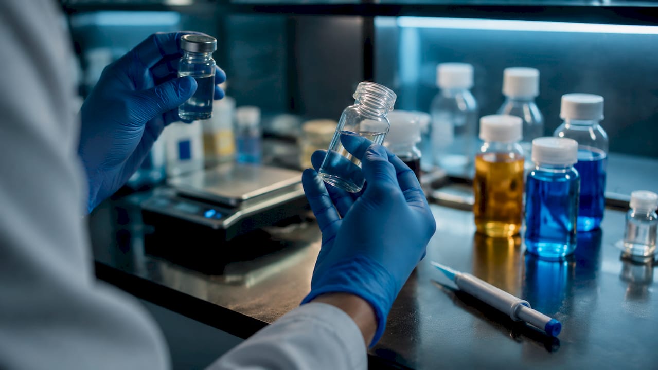
How to choose the right solvent for peptide reconstitution
Solvent selection is the decision with the highest impact on both peptide stability and downstream experimental validity. The wrong solvent can precipitate the peptide, alter its charge state, or introduce microbial contamination into a multi-use vial.
Solvent Best use case Key limitation Bacteriostatic water (0.9% benzyl alcohol) Multi-dose vials, peptides stable in neutral aqueous solution Not suitable for single-use protocols requiring preservative-free solvent Sterile water for injection Single-use reconstitution, sensitive peptides No preservative; discard within 24 hours after opening Dilute acetic acid (0.1–1%) Basic or hydrophobic peptides with poor aqueous solubility May require pH adjustment before use DMSO (dimethyl sulfoxide) Highly hydrophobic peptides resistant to aqueous solvents Risks oxidation of cysteine and methionine residues Normal saline (0.9% NaCl) Not recommended Ionic interactions destabilize peptides; no preservative
BAC water contains 0.9% benzyl alcohol, which inhibits bacterial growth after vial puncture. This makes it the standard choice for multi-dose research vials where the stopper will be punctured repeatedly over days or weeks. Sterile water is appropriate only for single-use scenarios, since it carries no preservative and supports microbial growth once opened.
For basic peptides with poor solubility in neutral water, dilute acetic acid (typically 0.1% to 1% v/v) protonates the peptide and improves dissolution. DMSO is reserved for highly hydrophobic sequences that resist aqueous reconstitution entirely, but researchers must account for the risk of oxidizing sensitive residues. Normal saline is generally avoided because ionic interactions between sodium chloride and charged peptide residues can destabilize the molecule, and saline lacks any preservative function for multi-dose use.
Pro Tip: When in doubt about solubility, consult the manufacturer’s certificate of analysis or synthesis report. The isoelectric point and net charge of the peptide sequence are the most reliable predictors of which solvent class will work without precipitation.
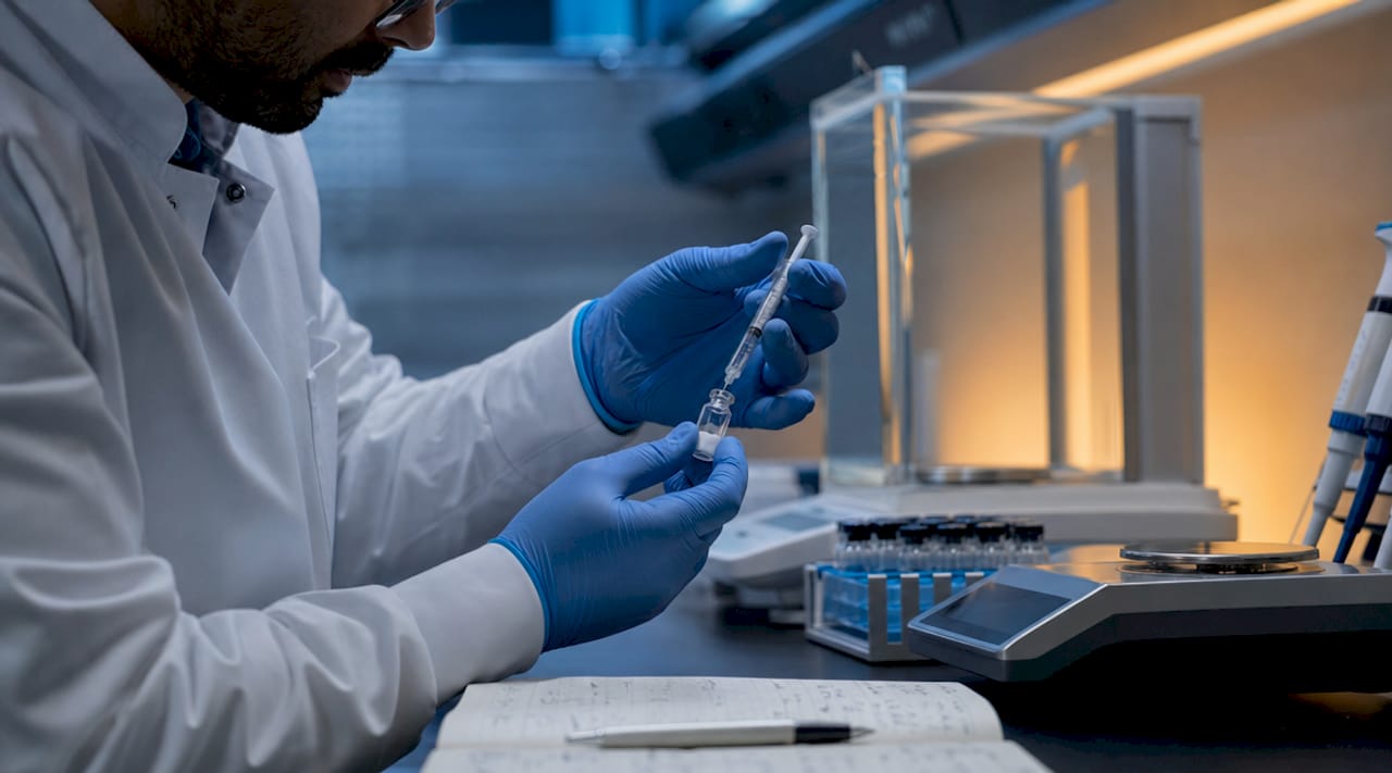
Step-by-step peptide reconstitution procedure
A reproducible reconstitution procedure eliminates the most common sources of variability in peptide research: condensation contamination, mechanical denaturation, and concentration error. Follow this sequence for every vial.
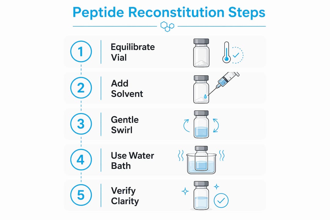
Equilibrate the vial to room temperature. Allow 15 to 20 minutes before opening. Cold vials generate condensation on the stopper surface, and that moisture can enter the vial during puncture, introducing contamination and diluting the peptide.
Prepare the work surface. Use a clean, low-particulate environment. Gather a 1 to 3 mL syringe with an 18 to 22 gauge needle for reconstitution and a separate U-100 insulin syringe with a 29 to 31 gauge needle for dosing. Using separate syringes for reconstitution and dosing prevents cross-contamination and concentration errors.
Disinfect the vial stopper. Wipe the rubber stopper with a 70% isopropyl alcohol swab and allow it to dry completely before puncture. A wet stopper introduces alcohol into the vial.
Calculate solvent volume. Determine the target concentration before drawing solvent (see the concentration section below). Use the peptide reconstitution calculator at PeptidesFromChina to verify volumes before proceeding.
Inject solvent slowly down the vial wall. Direct the needle tip toward the inner glass wall and depress the plunger slowly. Injecting solvent down the wall prevents mechanical disruption of the lyophilized cake through shear stress. Forceful direct injection onto the powder can denature peptide conformation.
Dissolve without shaking. Do not shake the vial. Shaking introduces air-liquid interfaces that denature peptides. Gently swirl or roll the vial between palms until the powder dissolves completely.
Verify solution clarity. A clear, colorless solution confirms successful reconstitution. Cloudiness, visible particles, or discoloration indicate instability, aggregation, or contamination. Do not use a compromised solution.
Label the vial immediately. Record peptide name, concentration, solvent type, and date of reconstitution. Unlabeled vials are a documented source of dosing errors in research labs and should never be returned to storage without complete annotation.
Pro Tip: If the powder does not dissolve within five minutes of gentle swirling, place the vial in a water bath at 37°C for two to three minutes, then continue swirling. Do not exceed 40°C, as elevated temperatures accelerate peptide degradation.
How to calculate peptide concentration and dosing volumes accurately
Concentration calculation is the step where most lab errors originate, and those errors propagate directly into experimental outcomes. The formula is straightforward: concentration (mg/mL) equals peptide mass (mg) divided by solvent volume (mL).
For a 5 mg peptide vial with 2 mL of BAC water added, the resulting concentration is 2.5 mg/mL. Targeting 2.5 to 5 mg/mL balances peptide stability with practical injection volumes when using insulin syringes for research dosing.
Key calculation principles:
Convert micrograms to milligrams before calculating (1,000 mcg = 1 mg) to avoid order-of-magnitude errors.
Target doses of 10 to 20 units on a 100-unit insulin syringe. Volumes below 5 units are difficult to measure accurately and introduce proportional dosing error.
For a U-100 insulin syringe, 1 unit equals 0.01 mL. A 10-unit draw equals 0.1 mL.
Adjust solvent volume to achieve a concentration where the intended dose falls within the 10 to 20 unit range on the syringe scale.
Peptide mass Solvent added Concentration 1 mg dose volume Insulin syringe units 5 mg 1 mL 5 mg/mL 0.2 mL 20 units 5 mg 2 mL 2.5 mg/mL 0.4 mL 40 units 10 mg 2 mL 5 mg/mL 0.2 mL 20 units 10 mg 4 mL 2.5 mg/mL 0.4 mL 40 units
External tools such as the Au Peptide Labs calculator provide an additional verification step for researchers who want to cross-check manual calculations before drawing solvent.
Proper storage practices and stability considerations after reconstitution
Post-reconstitution storage is where peptide integrity most commonly degrades in practice. The reconstituted solution is far more vulnerable than the lyophilized powder, and storage conditions directly determine how long the peptide remains usable.
Core storage guidelines:
Store reconstituted peptides at 2 to 8°C in the main body of the refrigerator, not the door. Door storage exposes vials to repeated temperature fluctuations with each opening.
BAC water-reconstituted peptides are stable for 2 to 4 weeks under proper refrigeration. Sterile water-reconstituted solutions should be discarded within 24 hours.
Never freeze a reconstituted solution. Freeze-thaw cycles promote aggregation and structural damage. If long-term storage is required, keep the peptide in lyophilized form and reconstitute only the volume needed.
Inspect the solution before each use. Discard any vial showing cloudiness, color change, or visible particulates, as these are signs of degradation or contamination.
Label every vial with the reconstitution date and planned discard date. A vial without a discard date will inevitably be used past its stability window.
The lyophilized powder, by contrast, tolerates room temperature storage for short periods and remains stable for one to two years at or below 4°C in a sealed, desiccated container. The transition from powder to solution is irreversible from a stability standpoint. Reconstitute only what will be used within the stability window of the chosen solvent.
Troubleshooting and handling difficult-to-dissolve peptides
Some peptides resist standard aqueous reconstitution regardless of technique. The causes are predictable: high hydrophobicity from aromatic or aliphatic residue clusters, strong aggregation tendencies from beta-sheet-forming sequences, or net charge near zero at physiological pH.
When standard reconstitution fails, apply this decision sequence:
Try dilute acetic acid (0.1% to 1%) for basic peptides, or dilute ammonium hydroxide (0.1%) for acidic peptides, before moving to organic solvents.
For hydrophobic peptides, use a two-step protocol with DMSO: dissolve the peptide in a small volume of DMSO first, then dilute slowly with aqueous solvent to the target concentration. DMSO volume should not exceed 10% of the final solution to avoid precipitation.
Avoid forceful injection and shaking at all stages. These introduce aggregation that cannot be reversed by further dissolution attempts.
If the solution remains cloudy after all solvent adjustments, the peptide may be degraded at the API level. Consult the batch certificate of analysis and contact the synthesis facility before proceeding.
Pro Tip: Sonication in a water bath sonicator for 30 to 60 seconds at room temperature can break up aggregates in difficult peptides without the thermal or mechanical damage caused by probe sonication or shaking.
Recognizing a degraded or improperly dissolved solution before use is as important as the reconstitution technique itself. A compromised solution produces unreliable data and wastes the entire batch.
Key takeaways
Successful peptide reconstitution requires precise solvent selection, controlled injection technique, accurate concentration calculation, and strict post-reconstitution storage within the stability window of the chosen solvent.
Point Details Equilibrate before opening Allow vials 15 to 20 minutes at room temperature to prevent condensation contamination. Match solvent to peptide Use BAC water for multi-dose vials; reserve DMSO for hydrophobic peptides with aqueous solubility failure. Inject slowly down the wall Direct solvent along the inner glass wall to prevent shear-induced denaturation of the lyophilized cake. Calculate for syringe precision Target 10 to 20 units on a U-100 insulin syringe to minimize volumetric dosing error. Label and discard on schedule BAC water solutions are stable 2 to 4 weeks at 2 to 8°C; sterile water solutions must be discarded within 24 hours.
What the protocol actually demands in practice
The written steps in any lyophilization protocol guide look straightforward. The execution is where researchers consistently lose reproducibility, and the failures are almost always procedural rather than chemical.
Vial equilibration is the step most frequently skipped under time pressure, and it is the one that introduces the most insidious contamination. Condensation on a cold stopper is invisible. It enters the vial silently and shows up later as cloudiness or unexpected biological activity in the assay. The 15-minute wait is not a formality.
The injection technique is similarly underestimated. Researchers who have worked with lyophilized biologics understand that the powder cake is structurally fragile. Directing solvent onto the cake rather than the glass wall is the equivalent of pressure-washing a sand castle. The mechanical disruption is real, and the resulting aggregates do not fully redissolve regardless of swirling time.
Concentration calculation errors are the most consequential failure mode because they are silent. A vial labeled at the wrong concentration produces data that looks clean but is systematically wrong. The calculation itself takes 30 seconds. Verifying it with a second tool, such as the PeptidesFromChina calculator or an external resource, takes another 30 seconds. That minute of verification is the difference between a reproducible experiment and a wasted batch.
Storage discipline is where even experienced labs cut corners. Reconstituted peptides stored in the refrigerator door, used past the four-week window, or frozen and thawed once “just to extend shelf life” are not the same peptides that came out of the lyophilizer. The stability window exists for a reason. Labeling vials with a discard date and actually discarding them on that date is the only practice that maintains data integrity across a multi-week experiment.
— Sam Levin
Source your peptides with the traceability the protocol requires
A reconstitution protocol is only as reliable as the peptide it starts with. Batch-to-batch inconsistency at the API level, undisclosed excipients, or poor lyophilization at the manufacturing stage all produce reconstitution failures that no technique can fix. PeptidesFromChina maintains direct relationships with established synthesis facilities and provides independent purity verification for each batch, so researchers can confirm what is in the vial before the protocol begins. Browse the research peptide catalog for lyophilized peptides with traceable batch documentation, and review the peptide quality sourcing page to understand how verification is conducted before any batch ships.
FAQ
What solvent is best for reconstituting most research peptides?
Bacteriostatic water containing 0.9% benzyl alcohol is the standard choice for multi-dose research vials because it inhibits bacterial growth after puncture and supports stable storage for 2 to 4 weeks at 2 to 8°C.
Why should you never shake a reconstituted peptide vial?
Shaking introduces air-liquid interfaces that denature peptides at the molecular level. Gentle swirling or rolling dissolves the powder without compromising the peptide’s native conformation.
How long is a reconstituted peptide stable in the refrigerator?
BAC water-reconstituted peptides are stable for 2 to 4 weeks at 2 to 8°C. Sterile water-reconstituted solutions carry no preservative and should be discarded within 24 hours of preparation.
What does a cloudy reconstituted solution indicate?
Cloudiness, visible particulates, or discoloration signal aggregation, instability, or contamination. A properly reconstituted peptide solution is clear and colorless; compromised solutions should not be used.
When should DMSO be used for peptide reconstitution?
DMSO is appropriate for highly hydrophobic peptides that fail to dissolve in aqueous solvents. It should be used in a two-step protocol, with DMSO volume kept below 10% of the final solution, and researchers must account for the risk of oxidizing cysteine or methionine residues.