How Peptide Quantification Methods Work for Researchers

Peptide quantification is defined as the systematic measurement of peptide concentrations in biological or synthetic samples using validated analytical techniques. The three primary method categories are liquid chromatography tandem mass spectrometry (LC-MS/MS), reversed-phase HPLC, and immunoassays such as ELISA. Each approach operates through a distinct detection principle, and selecting the wrong one for a given matrix introduces errors that propagate through every downstream calculation. Researchers in biochemistry and molecular biology need to understand how peptide quantification methods work before committing to a workflow, because method choice directly determines data reliability, sensitivity limits, and reproducibility across batches.
How peptide quantification methods work: LC-MS/MS as the primary approach
LC-MS/MS using the bottom-up proteomics workflow is the gold standard for measuring peptide concentrations in complex biological matrices. The workflow begins with enzymatic digestion, typically using trypsin, which cleaves proteins at lysine and arginine residues to generate peptide fragments of predictable mass and charge. Those fragments are then separated by liquid chromatography before entering the mass spectrometer for targeted detection.
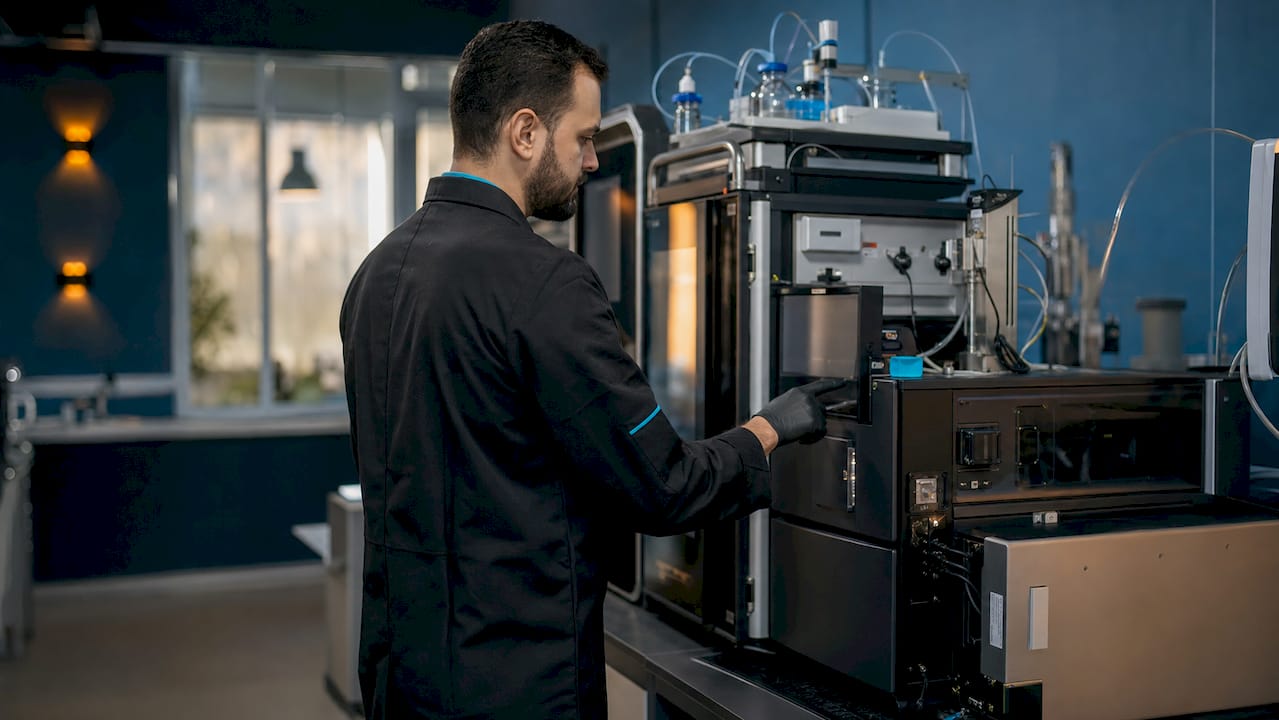
The detection step relies on Multiple Reaction Monitoring (MRM) using a triple quadrupole instrument. MRM selects a specific precursor ion in the first quadrupole, fragments it in the collision cell, and then detects a defined product ion in the third quadrupole. This two-stage selection gives MRM its specificity advantage over UV-based detection. MRM transitions achieve lower limits of quantification (LLOQ) between 0.60 and 29 nM on modern targeted platforms, which is sufficient for most biomarker and pharmacokinetic studies.
Matrix effects remain the most common source of error in LC-MS/MS quantification. Ion suppression from co-eluting matrix components can artificially reduce measured signal, causing underestimation of true peptide concentration. Two strategies correct for this:
Stable isotope-labeled internal standards (SIL-IS) co-elute with the analyte and experience identical suppression, so the analyte-to-SIL-IS ratio stays accurate.
Surrogate matrix calibration replaces the biological matrix with a surrogate fluid for calibrator preparation, reducing variability when authentic matrix is unavailable.
Pro Tip: Always verify that your SIL-IS co-elutes within 0.05 minutes of the native peptide. A retention time shift indicates incomplete isotope incorporation or a purity issue in the labeled standard itself.
The HPLC, LC-MS, and NMR analysis guide from PeptidesFromChina covers instrument configuration details relevant to researchers setting up these workflows from scratch.
How does reversed-phase HPLC contribute to peptide quantification accuracy?
Reversed-phase HPLC (RP-HPLC) with C18 stationary phases remains a cornerstone for peptide separation and purity assessment. The method separates peptides based on hydrophobic interactions between the analyte and the nonpolar stationary phase, with elution driven by increasing organic solvent concentration in the mobile phase. C8 and C4 phases are used for more hydrophobic peptides that bind too strongly to C18 under standard conditions.
Several parameters control resolution and peak shape in RP-HPLC method development:
Column pore size: 300 Å pores are standard for peptides above 3 kDa; 100 Å pores suit smaller fragments.
Particle morphology: sub-2-micron or core-shell particles reduce band broadening and improve peak efficiency.
Gradient slope: shallower gradients improve resolution of closely eluting impurities but increase run time.
Ion-pairing modifiers: trifluoroacetic acid (TFA) or heptafluorobutyric acid (HFBA) sharpen peaks for basic peptides.
UV detection at 214 nm measures peptide bond absorbance and provides a near-universal signal for all peptides. Detection at 280 nm is selective for tryptophan and tyrosine residues, which is useful when those residues are present and a more specific signal is needed.
Chromatography type Primary use Detection RP-HPLC (C18/C8) Purity and concentration UV 214 nm, MS Ion exchange (IEX) Charge variant profiling UV, conductivity Size exclusion (SEC) Aggregation and MW distribution UV 280 nm Hydrophobic interaction (HIC) Conformational variant separation UV
Orthogonal chromatography methods such as IEX and SEC do not replace RP-HPLC but complement it. SEC detects aggregates that RP-HPLC co-elutes with the monomer peak. IEX resolves deamidation variants that differ by charge but not hydrophobicity. Coupling any of these separations with mass spectrometry confirms peptide identity beyond retention time alone, which is a critical distinction when working with structurally similar analogs.
What are the main immunoassay formats for peptide quantification?
ELISA formats divide into two categories based on peptide size. Sandwich ELISA requires two distinct epitopes and works well for peptides above approximately 1.5 kDa. Competitive ELISA uses a single antibody and is the practical format for smaller peptides where two simultaneous antibody binding events are geometrically impossible.
Antibody cross-reactivity is the most common failure mode in peptide immunoassays. An antibody raised against a target peptide may bind structurally related sequences, metabolites, or truncated forms, inflating the measured concentration. This problem is particularly acute for peptide families with conserved sequence motifs, such as GLP-1 analogs or neuropeptides with shared C-terminal amidation.
The SISCAPA workflow addresses this limitation directly. SISCAPA combines immunoaffinity enrichment with mass spectrometry detection. Antibodies capture the target peptide from a complex matrix, concentrating it before LC-MS/MS analysis. The result is improved sensitivity and specificity compared to either method alone, with the structural confirmation that only MS can provide.
Amino acid analysis (AAA) offers an orthogonal route to absolute quantification. AAA hydrolyzes the peptide to its constituent amino acids and measures each by chromatography, providing a concentration value independent of antibody specificity or MS ionization efficiency. AAA is slower and more labor-intensive than LC-MS/MS, but it serves as a reference method for calibrator preparation and for validating SIL-IS stock concentrations.
Bioassays measure functional activity rather than mass concentration. They are not quantification methods in the strict analytical sense, but they provide a functional readout that mass-based methods cannot. A peptide that measures correctly by LC-MS/MS but shows no receptor activation in a bioassay has a structural defect that quantification alone would miss.
How do computational tools improve peptide quantification in complex datasets?
Label-free quantification (LFQ) and isobaric labeling represent two distinct computational strategies for measuring peptide concentrations across multiple samples. LFQ compares raw MS signal intensities between runs without chemical modification, which simplifies sample preparation but requires careful normalization to correct for run-to-run variation. Isobaric labeling with reagents such as TMT (tandem mass tags) multiplexes up to 18 samples in a single MS run, reducing instrument time but introducing ratio compression artifacts that require computational correction.
FragPipe and TMT-Integrator are the primary software platforms for isobaric-labeled proteomics data. TMT-Integrator normalizes quantitative data using ratio-to-reference and median centering approaches, then aggregates results across peptide, protein, and pathway levels. The platform handles multi-plex integration, which is critical when an experiment spans more batches than a single TMT kit can accommodate.
The GoldenHaystack algorithm addresses a specific gap in data-independent acquisition (DIA-MS) analysis. Standard DIA software assigns quantification only to peptides matched to a spectral library. GoldenHaystack quantifies unassigned ions by analyzing MS signal patterns across large datasets, recovering peptides that sequence-based methods miss entirely. The algorithm also runs faster than conventional tools like DIA-NN, which matters when processing large cohort studies.
Key computational steps that affect quantification reliability:
Missing value imputation: replacing absent signals with noise-level estimates or minimum observed values changes downstream statistical outcomes.
Batch effect correction: median centering within each batch removes systematic shifts introduced by reagent lot changes or instrument drift.
Normalization strategy: ratio-to-reference normalization anchors each sample to a common reference channel, making cross-batch comparisons valid.
Protein rollup: summing or averaging peptide-level intensities to the protein level requires decisions about which peptides to include and how to weight them.
Pro Tip: Run a principal component analysis (PCA) on your normalized peptide matrix before any biological comparison. Samples clustering by batch rather than by biological group signal a normalization failure that will invalidate your results.
Researchers working on peptide biomarker studies will find that computational normalization choices often have a larger effect on final results than instrument platform selection.
Key Takeaways
Accurate peptide quantification requires combining targeted detection methods like LC-MS/MS with orthogonal verification techniques and rigorous computational normalization to produce reproducible concentration data.
Point Details LC-MS/MS is the primary method MRM on triple quadrupole instruments achieves LLOQ between 0.60 and 29 nM in complex matrices. Matrix effects require active correction Stable isotope-labeled internal standards and surrogate matrix calibration prevent ion suppression errors. Immunoassays carry cross-reactivity risk SISCAPA hybrid workflows combine antibody enrichment with MS detection to improve specificity. Computational normalization is not optional Batch effects from multi-plex experiments require ratio-to-reference or median centering to produce valid comparisons. Orthogonal methods confirm identity Combining RP-HPLC, MS/MS fragmentation, and amino acid analysis is required to verify peptide sameness and purity.
What I’ve learned from watching researchers pick the wrong method first
The most consistent mistake I see in peptide quantification workflows is treating method selection as a secondary decision. Researchers often choose a platform based on what is available in their facility rather than what the sample matrix and target concentration range actually demand. A sandwich ELISA that works well for a 5 kDa neuropeptide in cerebrospinal fluid will fail completely when applied to a 900 Da synthetic analog in plasma, not because the researcher made a technical error, but because the format was never appropriate for the analyte.
The second pattern worth naming is over-reliance on a single method for both identity confirmation and quantification. RP-HPLC purity data tells you what fraction of the signal comes from the main peak. It does not tell you whether that peak is the correct peptide or a closely eluting impurity with similar hydrophobicity. Orthogonal analytical confirmation using MS/MS fragmentation is not a redundant step. It is the step that separates a measurement from a guess.
Computational tools have genuinely changed what is possible in multiplexed quantification studies. But they also introduce a new failure mode: researchers who trust normalized output without auditing the normalization itself. A PCA plot takes five minutes to generate and immediately reveals whether batch effects survived the correction step. Skipping it is a reproducibility risk that shows up only when someone tries to replicate the experiment.
The practical recommendation is a three-layer strategy: use LC-MS/MS as the primary quantification method, apply at least one orthogonal technique for identity verification, and audit computational normalization before interpreting biological results. This is not a conservative approach. It is the minimum required to produce data that holds up under scrutiny.
— Sam Levin
Research-grade peptides from PeptidesFromChina for quantification studies
Quantification workflows depend on the quality of the starting material. A peptide with undisclosed impurities or inconsistent batch composition introduces variability that no analytical method can fully correct.

PeptidesFromChina supplies research-grade peptides with manufacturer-side analytical documentation that may be available depending on the product and batch, giving researchers a documented baseline before any quantification experiment begins. The catalog covers a broad range of research targets, from signaling peptides to GLP-1 analogs, with batch traceability that supports reproducibility across study phases. PeptidesFromChina coordinates sourcing from established Chinese manufacturing facilities together with international logistics without relying on anonymous retail-reseller supply chains. Researchers who need well-characterized starting material for LC-MS/MS or immunoassay workflows will find the sourcing documentation directly relevant to their validation requirements.
FAQ
What is the most sensitive peptide quantification method?
LC-MS/MS using MRM on a triple quadrupole instrument achieves the lowest detection limits, with LLOQ values between 0.60 and 29 nM in complex biological matrices. SISCAPA workflows extend sensitivity further by combining immunoaffinity enrichment with MS detection.
How does RP-HPLC differ from LC-MS/MS for peptide measurement?
RP-HPLC measures UV absorbance at 214 nm and provides purity data but cannot distinguish isobaric or isomeric peptides. LC-MS/MS adds mass-based identity confirmation and achieves lower detection limits, making it the preferred method for quantification in complex samples.
Why do matrix effects matter in peptide quantification?
Co-eluting matrix components suppress ionization in the mass spectrometer, causing the instrument to underreport true peptide concentration. Stable isotope-labeled internal standards and surrogate matrix calibration correct for this suppression by normalizing the analyte signal to a co-eluting reference.
When should researchers use competitive ELISA instead of sandwich ELISA?
Competitive ELISA is the correct format for peptides below approximately 1.5 kDa, where the analyte is too small to accommodate two antibodies simultaneously. Sandwich ELISA requires two distinct epitopes and is appropriate only for larger peptides.
What does the GoldenHaystack algorithm do that standard DIA software cannot?
GoldenHaystack quantifies peptides that lack spectral library matches by analyzing unassigned MS ion patterns across large datasets. Standard DIA tools skip these ions entirely, so GoldenHaystack recovers quantification data that conventional sequence-based methods miss.